Types of breast calcification
Macrocalcifications
Microcalcifications are more extensive calcium deposits that are often benign and connected with noncancerous conditions such as cysts, areas of benign breast tissue changes, and fibroadenomas (benign breast tumors). According to St. Vincent’s Private Hospital, macrocalcifications often show up in 50% of women that are 50 years old. While they also appear in 10% of women under 50 years old.
Microcalcifications
On the other hand, microcalcifications are tiny calcium deposits that are tinier than 1/50th of an inch (1 mm) in size. These are more closely assessed since they can be an early sign of potential abnormalities, including precancerous or cancerous changes in the breast tissue. The characteristics, pattern, and distribution of microcalcifications can help radiologists in terms of determining the possibility for further investigation.
Causes of breast calcification
Radiations
The involvement of high-energy radiation, known as radiation therapy, which is used to treat cancer, can cause breast calcifications. According to Breast Cancer.org, as a result of radiation or surgery to the chest area, calcifications with fat necrosis can happen. Over time, radiation therapy can cause changes in the breast tissue.
The radiation can lead to various effects, including changes in blood vessels, fibrosis (scarring), and the formation of calcifications or calcium deposits. This is because the radiation can damage the structures and cells within the breast. However, it’s important to note that the calcifications are generally considered benign when they develop due to radiation therapy. This means they’re not cancerous, but these calcifications require careful evaluation and monitoring.
Breast infection or mastitis

According to Beverly Hospital, calcifications can be associated with mastitis (infection), fibrocystic breast changes, and injuries. Therefore, they can present normal occurrences in breast tissues. It’s important to know that mastitis commonly occurs in lactating women, specifically when breastfeeding. But this infection can also affect women even if they’re not breastfeeding. When mastitis happens, it often leads to redness, swelling, pain in the affected breast, and warmth.
Additionally, the inflammation may result in the formation of scar tissue since it can cause damage to the breast tissue. Sometimes, this scar tissue can result in the development of breast calcifications when they calcify over time. In rare cases, calcification associated with an infection can be related to a more serious condition, such as chronic inflammation.
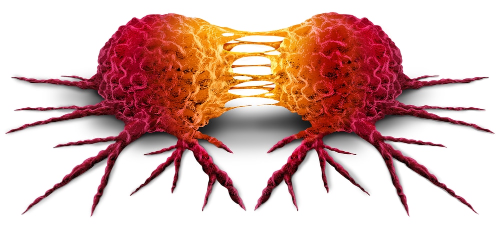
Breast cancer

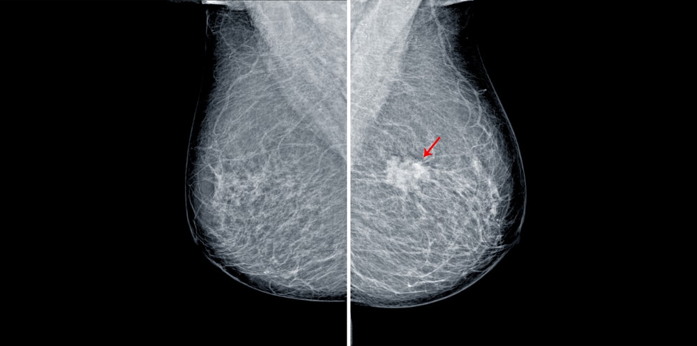
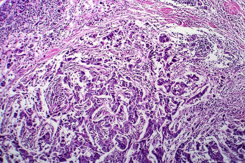
According to Cancer Treatment Centers of America, despite breast calcifications being reasonably common, they can be the earliest sign of breast cancer. Ductal carcinoma in situ (DCIS) is a specific type of breast cancer that is usually associated with the presence of microcalcifications. A non-invasive form of breast cancer like DCIS is when abnormal cells have not spread into surrounding breast tissue when they’re confined to the milk ducts.
Furthermore, when they calcify, these abnormal cells can form tiny specks or clusters of calcium, known as microcalcifications. These microcalcifications are an important finding for the early detection of DCIS since they appear as white dots or spots on a mammogram. The presence of particular characteristics in microcalcifications can prompt further evaluation and raise concern, even though not all breast calcifications indicate cancer.
Fat necrosis

Fat necrosis can be a potential cause of breast calcification. In a study published by the National Institutes of Health, fat necrosis manifests as firmness and induration on gross pathology. This is because fat necrosis first appears as a hemorrhage area in fat. Saponification occurs subsequently, where the lesion may become yellow. Calcification presents as a chalky white lesion and a yellow-gray mass called fibrosis.
Moreover, fat necrosis is an inflammatory and sterile process that results from aseptic saponification of tissue lipase and fat utilizing blood. This condition can happen due to surgery, trauma, injury, or even without any evident cause. The damaged fat cells can release calcium when fat necrosis occurs, leading to calcification formation within the breast tissue.
Intraductal papilloma
Intraductal papilloma can be a potential cause of breast calcification. A study published in the National Library of Medicine showed that intraductal papilloma can be found within breast ducts as a benign tumor. What causes tumor growth is the abnormal proliferation of ductal epithelial cells. What’s usually found centrally posterior to the nipple is a solitary intraductal papilloma, which affects the central duct.
Moreover, multiple intraductal papillomas can be found in any breast quadrant since they’re located peripherally. This affects the peripheral ducts. Keep in mind that intraductal papilloma usually appears as a small finger-like or wart-like projection, which can cause symptoms like palpable lump or nipple discharge.
Fibrocystic changes
According to the MD Anderson Cancer Center, fibrocystic changes are rope-like in texture and can feel lumpy. They’re often associated with calcifications. They’re noncancerous or benign breast tissue changes, often leading to cysts or lumps formation. The calcifications don’t indicate breast cancer when associated with fibrocystic changes. But the characteristics and appearance of calcifications in imaging studies can sometimes be a resemblance of those seen in baleful breast lesions.
This is why careful monitoring and evaluation of these calcifications are important to distinguish them from findings that are potentially suspicious. A healthcare professional or a breast specialist can review the mammogram or other imaging results when breast calcifications are detected in the context of fibrocystic changes. These professionals can assess the calcifications through their nature and characteristics.
Mammary duct ectasia

This refers to the abnormal widening of breast ducts in 3 mm at the ampulla or greater than 2 mm diameter, according to Radiopaedia. Mammary duct ectasia is usually found in females whose ages range from 50-60 years. It can occasionally be seen in children, while it’s rare for males.
It’s important to take note that mammary duct ectasia can cause discomfort and inflammation in the nipple area. The ducts can be filled with debris or become dilated when inflammation occurs in the milk ducts. This debris can result in the development of calcifications within the affected ducts when they include calcium deposits over time.
Trauma or injury

Calcifications can occur due to injuries to the breast caused by auto accidents or falls, according to Compass Ontology. Therefore, trauma or injury can be a potential cause of breast calcifications. According to Pure Mammography, the result of breast trauma can be troubling to the person, even though most breast injuries don’t result in permanent damage to the breasts. If you currently experience breast trauma, symptoms such as bruising, lump in the breast, and pain can convince you that you have breast cancer.
However, keep in mind that these tissue injuries most commonly require time for recovery, despite being uncomfortable with it. In terms of the body’s healing process, it may involve scar tissue formation in response to the injury. This scar tissue can undergo calcification in some cases.
Aging

This is considered to be a common factor associated with the development of breast calcifications. Changes occur within the breast tissue as women age, including calcifications and their formation. These calcifications appear as small white spots or dots when they’re detected on mammograms.
Medical News Today confirmed that breast calcification is connected to advanced aging. This leads to a higher possibility of changes in noncancerous breast cells. These can also leave behind calcium deposits. It’s believed that breast tissue enters natural aging processes, like the deposition of fibrous tissue and the involution (the shrinking of glandular tissue).
Breast arterial calcification

This refers to the hardening or calcification of the arteries within the breast tissue. Breast arterial calcification usually occurs due to the buildup of calcium deposits in the blood vessels and the result of age-related changes. According to the American Heart Association, breast arterial calcification is related to high blood pressure, inflammation, aging, and Type 2 diabetes. This is also a marker of stiffening in the arteries.
Remember that this cause is often observed in older women and is often detected incidentally on mammograms. Along the course of the blood vessels in the breast, the calcifications appear as branching or linear patterns of calcified deposits. Moreover, breast arterial calcifications are often considered a typical age-related change that’s the same as arterial calcifications, which can occur in some parts of the body.
Symptoms of breast calcification
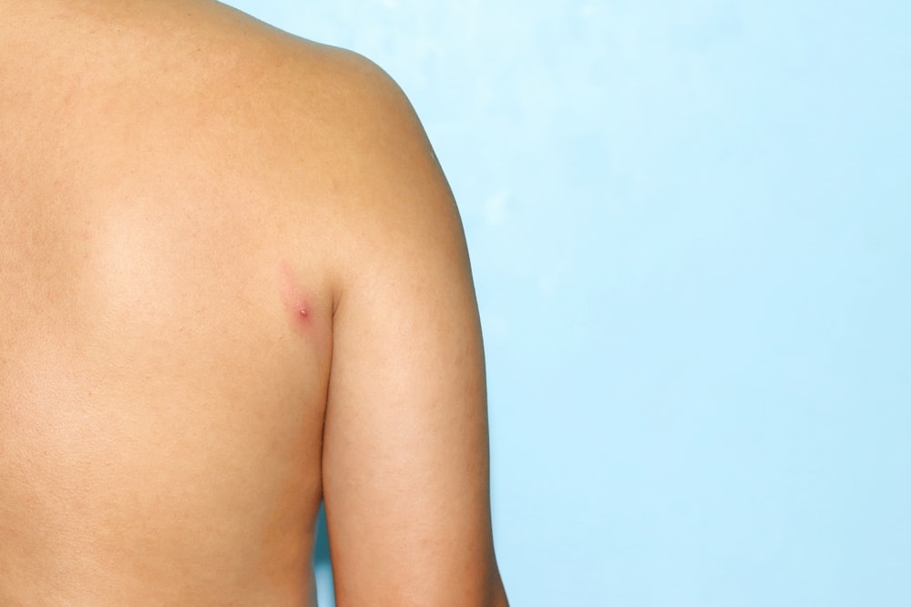
It’s important to note that breast calcifications don’t typically have symptoms since they’re too small to be felt. However, it’s still important to consult your doctor if you’re experiencing odd breast symptoms that can show an underlying condition associated with an infection or cancer. According to Healthline, potential symptoms include red or inflamed skin, lumps or bumps around your underarm or breast, nipple discharge or changes such as inversion, chronically itchy skin, and breast skin that’s puckered, scaly, or dimpled.
Treatments for breast calcification
Supportive care and education

They can be vital in managing any underlying concerns or conditions connected with breast calcifications. Supportive care and education can provide individuals with information about breast calcifications. This includes their causes, benign nature, and the associated conditions. This helps provide reassurance and alleviate concerns that most calcifications aren’t cancerous.
It’s normal for a patient to experience anxiety or worry when a diagnosis of breast calcifications takes place. Supportive care aims to provide counseling, emotional support, or access to support groups where individuals can find comfort in connecting with others who have similar concerns so they can share their experiences as well. Moreover, supportive care may involve recommendations for pain management techniques if this condi
tion is associated with symptoms such as discomfort or breast pain. These pain management techniques include warm compresses, over-the-counter pain medications, or supportive bra fittings.
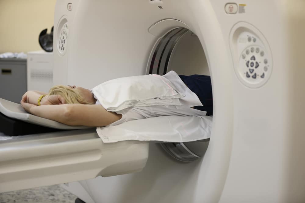
Surveillance and regular screening

These are important components of preventive care and ongoing monitoring for breast health. Surveillance and regular screening involve the regular and systematic monitoring of breast tissue. This includes the evaluation and detection of breast calcifications and other abnormalities. This is typically achieved with a standard screening tool known as mammography, which is appropriate for breast cancer.
The purpose of these components is to detect any developments or changes in breast tissue, including the presence of alterations or new calcifications in existing ones. Early intervention and treatment can be initiated by detecting any concerning findings, such as suspicious calcifications associated with breast cancer, in order to result in better outcomes.
Radiation therapy
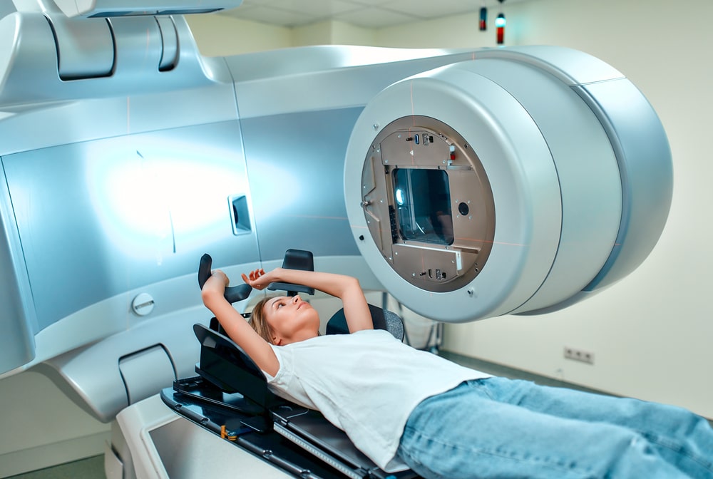
The treatment approach of radiation therapy for breast calcification is usually focused on follow-up examinations and regular monitoring. This may involve periodic imaging studies such as mammograms or other imaging modalities to assess the changes or stability in the calcifications over time.
It’s important to keep in mind that there are other concerning findings connected with breast calcifications, such as suspicious features on imaging, interventions, or additional tests, as well as changes in breast tissue to manage and evaluate the situation effectively. It’s always advisable to consult with a breast specialist or your healthcare provider if you have concerns about breast calcifications related to previous radiation therapy.
Hormonal therapy

This type of therapy can be utilized to manage underlying conditions associated with breast calcifications, such as fibrocystic changes or hormonal imbalances. Hormonal therapy involves the use of medications that influence hormone levels in the body. It can help reduce the symptoms associated with hormonal conditions, minimize the risk of the development of calcifications or further changes, and regulate hormonal imbalances.
Hormonal therapy may be recommended to help manage symptoms such as tenderness, cysts, or breast pain. This can be associated in the case of fibrocystic changes, which can contribute to the formation of calcifications. Hormonal therapy options may include selective estrogen receptor modulators (SERMs) like aromatase inhibitors or tamoxifen.
Observation and monitoring

These are important aspects of breast calcification management, specifically when they’re determined to be benign and not associated with underlying conditions. Observation and monitoring involve a proactive approach to track any changes in the calcifications over time, despite not being considered treatments in the traditional sense. A study published in the National Library of Medicine shows that improving the detection of malignant calcifications as early signs of cancer is important.
This is because the majority of lesions aren’t recalled immediately with detectable calcifications but are detected as interval cancer or are invasive at the time of diagnosis in the next screening round. By stability assessment, healthcare providers can assess whether the calcifications remain stable over time, including their benign nature, by comparing imaging studies or mammograms taken at different time points.
Lifestyle modifications

According to Acko, it’s important to adopt a healthy lifestyle for breast calcifications. This can include having a balanced diet, regular exercise, avoidance of smoking, and limited alcohol consumption. These habits can place large contributions to overall breast health.
After menopause, excess body weight is associated with an increased risk of breast cancer and other breast-related conditions. Regular physical activity like strength training, aerobic exercises, or activities like jogging or brisk walking can positively affect breast health. Exercise helps support overall well-being, maintains a healthy weight, and improves circulation.
Pain management

Keep in mind that rather than directly treating the calcifications themselves, the main focus of pain management is to alleviate the symptoms. This is because calcifications don’t require specific treatment and are typically benign. Naproxen sodium or ibuprofen are nonsteroidal anti-inflammatory drugs (NSAIDs) that can help reduce inflammation and pain associated with breast calcifications.
In some cases, individuals find ice packs or cold compresses helpful in reducing pain or swelling and numbing the affected area. To avoid direct contact with ice and to protect the skin, it’s important to wrap the cold compress in a cloth or towel. Moreover, topical creams that contain ingredients such as lidocaine or menthol may avoid temporary relief from discomfort or localized pain.
Treatment of underlying conditions

According to Medical News Today, if the calcifications are associated with another medical condition or may indicate breast cancer, the person will require treatment to kill the cancerous cells and to stop the spread of cancer. Take note that the type of treatment a person needs depends on the breast cancer type or its size and stage. Some potential treatments for breast cancer include radiation, surgery, hormone therapy, and chemotherapy.
On the other hand, conditions like intraductal papillomas are often removed surgically to alleviate symptoms, rule out any potential associated malignancy, and confirm the diagnosis. The surgical procedure may involve removing a small portion of breast tissue containing the papilloma or removing the affected duct.
Biopsy or tissue sampling

These may be recommended as a diagnostic procedure to further evaluate the underlying cause and nature of the calcifications. According to Cancer Treatment Centers of America, a test called a biopsy can identify the makeup of breast calcifications and their cells if they seem suspicious to the patient. Take note that core needle biopsy is the most common type of breast biopsy.
Your doctor places a hollow needle during a core needle biopsy on your breast in order to remove tissue samples from the suspicious area. On the other hand, a fine needle aspiration biopsy (FNAB) is a thin needle that’s used to extract cell samples from the area of concern for examination.
Surgical excision

According to St. Vincent’s Private Hospital, your doctor may sometimes recommend surgery to remove the calcification area from the breast. This is usually applied due to an unsuccessful needle core biopsy in terms of removing enough calcification or when there’s an indefinite result. A preoperative hook wire localization technique is utilized for the surgeon to pinpoint the area.
The preoperative hookwire localization breast surgery is divided into two parts: (1) before the operation, a hookwire is inserted under local anesthetic; and (2) during an operation, a calcification is removed under general anesthetic. Additionally, surgical excision may be advised to exclude the possibility of malignancy and to obtain a definitive diagnosis. This is advised if the calcifications demonstrate concerning features on imaging like clustering, architectural distortion, or suspicious morphology.